Biological materials

Biophysical Journal Volume 94 June 2008 4220-4232
Hierarchical Modeling of the Elastic Properties of Bone at Submicron Scales: The Role of Extrafibrillar Mineralization
Svetoslav Nikolov and Dierk Raabe
Max-Planck-Institut für Eisenforschung, Department of Microstructure Physics, Düsseldorf, Germany
Biophysical Journal 94(11) 4220–4232 Sim[...]
PDF-Dokument [872.3 KB]
 Biophysical Journal Volume 94 June 2008 4220-4232 Hierarchical Modeling of the Elastic Properties of Bone at Submicron Scales: The Role of Extrafibrillar Mineralization Svetoslav Nikolov and Dierk Raabe Max-Planck-Institut für Eisenforschung
The remarkable mechanical properties of bone related to its low density are essentially due to the bone’s complex, hierarchical microstructure from the macro- down to the nanoscale. At
length scales below several microns,
the diversity of bone tissues is reduced to different arrangements of mineralized collagen fibrils formed through self-assembly of soft collagen molecules and hard
mineral nanoparticles. At this level of organization, the effective mechanical properties of bone depend on the properties of the fibrils’ constituents, the fibrils’
microstructure, and orientation distribution, as well as on the mineral content
and the shape of the mineral particles. Clearly, the development of reliable structure-properties relations for mineralized collagen fibrils and for fibril arrays incorporating these
dependencies is of crucial importance, not only for the evaluation of the mechanical properties of bone, but also for better understanding of how the governing material-design
principles, growth processes, or diseased states influence the mechanical properties at submicron length scales. Such relations can also help answer some still unclear questions about
the bone structure and serve as a tool for designing of new bone implants and bioinspired nanocomposites.
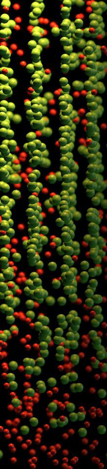
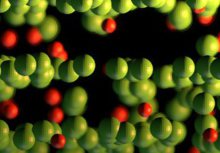
At the nanoscale, bone consists of:
Collagen type I molecules (triple helices ;300 nm long and 1.5 nm in diameter), self-assembled in a staggered fashion to form collagen fibrils with diameter of 100
nm.
Biological hydroxyapatite (HA) minerals with hexagonal unit cells.
Water, part of which provides hydrogen (H) bonds for both the collagen molecules and the collagen-mineral composite.
A relatively limited amount of noncollagenous proteins (NCPs) like the extrafibrillar proteins that glue together adjacent collagen fibrils.
It is now well established that the HA crystals inside the collagen fibrils grow primarily in the gaps between subsequent collagen molecules and are shaped as platelets with
typical average dimensions of ;50 3 25 3 3 nm, although
their length can vary from 15 to 150 nm, their width from 10 to 80 nm, and their thickness from 2 to 5 nm. The longest dimension of the HA platelets coincides with the c axis of
the HAcrystal unit cell and is oriented along the fibril axis. The
structure and the relative fraction of the extrafibrillar minerals are lesswell understood and are stillamatter of debate.Recent study by atomic force microscopy (AFM) confirmed
the existence of mineral-containing blobs on the fibrils surface. Hassenkam et al. and Kindt et al. obtained a more detailed picture of the extrafibrillar minerals via
high-resolution AFM imaging. They observed that each collagen fibril is individually coated with extrafibrillar HA minerals with various shapes and sizes and part of these mineral
formations are arranged
with a period of 67 nm, the same as that of the underlying microstructure of the naked collagen fibrils.
Using these results, in this project we modeled the elastic properties of bone at the level of mineralized collagen fibrils via step-by-step homogenization
from the staggered arrangement of collagen molecules up to an array of parallel mineralized fibrils. A new model for extrafibrillar mineralization is proposed, assuming that the
extrafibrillar minerals are mechanically equivalent to reinforcing rings coating each individual fibril. Our modeling suggests that no more than 30% of the total mineral content is
extrafibrillar and the fraction of
extrafibrillar minerals grows linearly with the overall degree of mineralization. It is shown that the extrafibrillar mineralization considerably reinforces the fibrils’ mechanical properties in
the transverse directions and the fibrils’ shear moduli. The model predictions for the elastic moduli and constants are found to be in a good agreement with the experimental data reported in
the literature.
Journal of Structural Biology 178 (2012) 290–299
SharkTeeth_JStructBiol_2012.pdf
PDF-Dokument [1.9 MB]
Journal of Structural Biology 178 (2012) 290-299
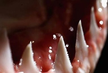
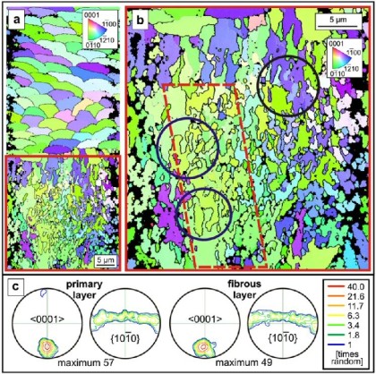
The teeth of two different shark species (Isurus oxyrinchus and Galeocerdo cuvier) and a geological fluoroapatite single crystal were
structurally and chemically characterized. In contrast to dentin, enameloid showed sharp diffraction peaks which indicated a high crystallinity of the
enameloid. The lattice parameters of enameloid were close to those of the geological fluoroapatite single crystal. The inorganic part of shark teeth consisted of fluoroapatite with a
fluoride content in the enameloid of 3.1 wt.%, i.e., close to the fluoride content of the geological fluoroapatite single crystal (3.64 wt.%). Scanning electron micrographs showed that the
crystals in enameloid were highly ordered with a special topological orientation (perpendicular towards the outside surface and parallel towards the center). By thermogravimetry, water,
organic matrix, and biomineral in dentin and enameloid of both shark species were determined. Dentin had a higher content of water, organic matrix, and carbonate than enameloid but contained
less fluoride. Nanoindentation and Vicker’s microhardness tests showed that the enameloid of the shark teeth was
approximately six times harder than the dentin. The hardness of shark teeth and human teeth was comparable, both for dentin and enamel/enameloid. In contrast, the geological fluoroapatite single
crystal was much harder than both kinds of teeth due to the absence of an organic matrix. In summary, the different biological functions of the shark teeth (‘‘tearing’’ for Isurus and
‘‘cutting’’ for Galeocerdo) are controlled by the different geometry and not by the chemical or crystallographic composition.
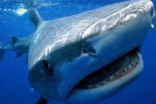 Tiger shark: Galeocerdo cuvier
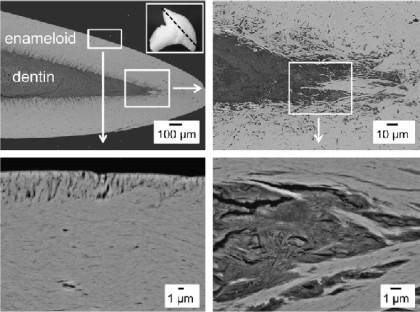 Scanning electron micrographs of an axial cross section-polished surface (see insert) of a tooth of Galeocerdo cuvier, showing the dentin-enameloid junction (backscattered electron mode).
Biological materials are hierarchically structured nanocomposites optimized through evolution to perform vital functions within the specific ecophysiological strains of living organisms. Most biological materials with structural functions consist of an organic matrix of structural biopolymers like collagen, chitin or cellulose which is modified and reinforced with various other proteins and in many cases also with biominerals. The most prominent examples like the bones of vertebrates, the exoskeletons of arthropods, and mollusk shells are known to possess excellent mechanical properties (e.g., in terms of stiffness-to-density ratio and fracture toughness) which are generated by structural and chemical alterations at different levels of their structural hierarchy. The origins of these properties, particularly the underlying structure-composition property relations, are the research subject in this field..
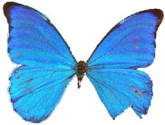
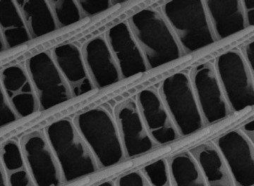 Photonic crystal of a butterfly wing
Acta Materialia 53 (2005) 4281-4292
The crustacean exoskeleton is an example of a structurally and mechanically graded biological nanocomposite
D. Raabe, C. Sachs, P. Romano
Acta Materialia 53 (2005) 4281–4292 biol[...]
PDF-Dokument [908.1 KB]
 Acta Materialia 53 (2005) 4281-4292 The crustacean exoskeleton is an example of a structurally and mechanically graded biological nanocomposite D. Raabe, C. Sachs, P. Romano
 Acta Materialia 53 (2005) 4281-4292 The crustacean exoskeleton is an example of a structurally and mechanically graded biological nanocomposite D. Raabe, C. Sachs, P. Romano
Acta Materialia 53 (2005) 4281-4292
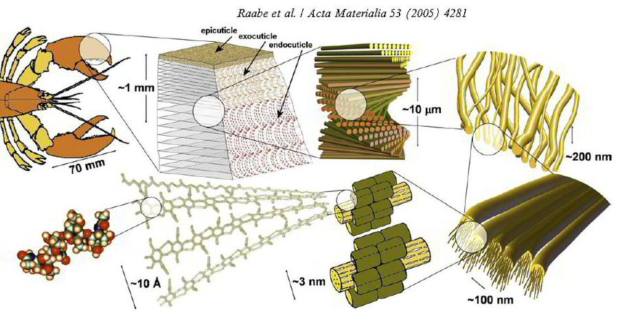
The exoskeleton material of arthropods consists of mineralized fibrous chitin-based tissue. The most characteristic feature of this biological nanocomposite material is its strictly hierarchical organization which reveals various structural levels: at the molecular level is the polysaccharide chitin itself. Its antiparallel alignment forms a-chitin crystals. The next structure
level is the arrangement of 18–25 of such molecules in the form of narrow and long crystalline units, which are wrapped by proteins, forming nanofibrils of about 2–5 nm diameter and
about 300 nm length. The next step in the scale consists in
the clustering of some of these nanofibrils into long chitin–protein fibers of about 50–300 nm diameter. These chitin–protein fibers form a planar woven and
periodically branched network (chitin–protein layers). The spacing between the fibers is filled up with proteins and biominerals of microscopic and nanoscopic size. The minerals are mostly
in the form of crystalline CaCO3, but amorphous particles may also occur depending on the species and molt cycle. The most characteristic level in the overall hierarchy, visible even
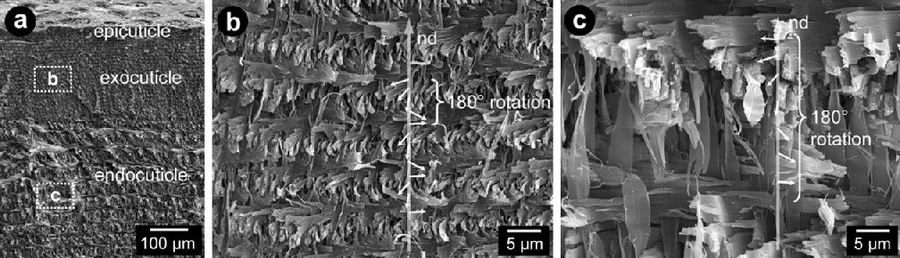
in an optical microscope, is referred to as a twisted plywood or Bouligand pattern. This structure is
formed
by the helicoidal stacking sequence of the fibrous chitin-protein layers. The thickness of one such twisted plywood or Bouligand layer corresponds to a
certain stacking density of planes which are gradually rotated about their normal axis, thereby creating complex structures which appear as mesoscale arches when cut in cross
sections. This study presents observations, which substantiate
that this structural picture of the exoskeleton material of arthropods must be completed by an additional feature, namely, by the occurrence of a pronounced mesoscopical structural
gradient of the twisted plywood pattern through the thickness of the exoskeleton material. Corresponding microindentation tests which were carried out through the cuticle thickness
further suggest that the structure gradients observed play an important role for the micromechanical design strategy inherent to such materials. The experiments are conducted on
the cuticle of the lobster Homarus americanus. This animal is a large arthropod (joint-limb animal) which belongs to the class of the crustaceans and the order of the
decapods. Its cuticle consists, like that of most arthropods, of the three main layers, epicuticle, exocuticle, and endocuticle, which are secreted by a single layer of epidermis
cells. The epicuticle (outer skin) is a very thin and waxy functional layer, which acts as a diffusion barrier to the surrounding. The exocuticle and endocuticle layers,
which carry the mechanical loads, consist of a hard mineralized fibrous chitin-protein tissue as described above.
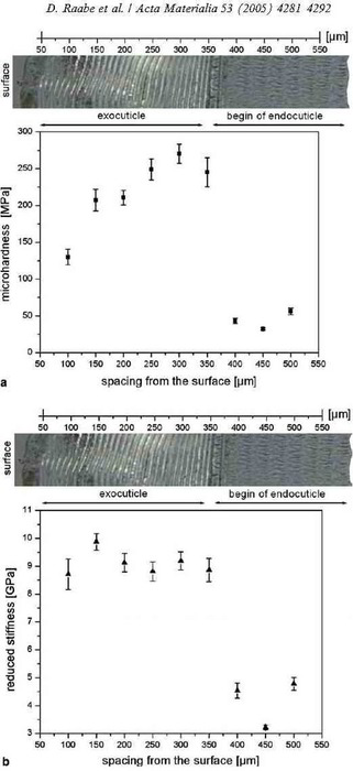
This paper presents an experimental study on the mechanical and structural gradients through the cuticle of Homarus americanus
(American lobster). The exocuticle (outer layer) is characterized by a very fine woven structure of the fibrous chitin-protein matrix
(twisted plywood structure) and by a high stiffness (8.5–9.5 GPa). The hardness increases within the exocuticle between the surface region (130 MPa) and
the region close to the interface to the endocuticle (270 MPa). In the endocuticle, which is characterized by a much coarser twisted plywood structure, both the stiffness (3–4.5 GPa) and
hardness (30–55 MPa) are much smaller than in the exocuticle. The transition in mechanical properties and structure between the exocuticle and endocuticle is abrupt. The differences underline
the important role of the internal structure of the twisted plywood structure and of the interface between the two cuticle layers for the overall
mechanical behavior of the exoskeleton. The excellent mechanical stability of the interface (irrespective of the change in the mechanical
properties) is attributed to the fact that the structural change of the twisted plywood pattern across the interface consists only of a change of the stacking density of the
chitin–protein layers. The observed gradients in stiffness and hardness
through the cuticle thickness are interpreted in terms of honeycomb mechanics of the twisted plywood structure. The possible role of gradients in protein cross-linking and in
the mineral content is also discussed.
Thermochimica Acta 463 (2007) 65-68
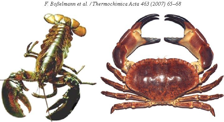
The composition of the exoskeleton of two crustacea: The American lobster Homarus americanus and the edible crab Cancer pagurus
F. Boßelmann, Romano, H. Fabritius, D. Raabe, M. Epple
Thermochimica Acta 463 (2007) 65–68 comp[...]
PDF-Dokument [300.8 KB]
 Thermochimica Acta 463 (2007) 65-68 The composition of the exoskeleton of two crustacea: The American lobster Homarus americanus and the edible crab Cancer pagurus F. Boßelmann, Romano, H. Fabritius, D. Raabe, M. Epple
 Thermochimica Acta 463 (2007) 65-68 The composition of the exoskeleton of two crustacea: The American lobster Homarus americanus and the edible crab Cancer pagurus F. Boßelmann, Romano, H. Fabritius, D. Raabe, M. Epple
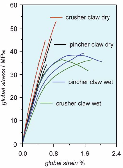
Crustaceans constitute a widespread class of organisms of both marine and land-dwelling species. They possess an exoskeleton [1] that stabilizes the whole body of the animal and
also serves for protection against predators. In the case
of lobsters and crabs, the claws are part of the exoskeleton optimized to serve as a cutting tool. The exoskeleton of crustaceans [2] is a biomineralized [3–6] structure which
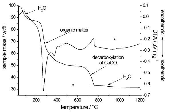
consists of an organic matrix (in this case -chitin) together with an inorganic mineral, in this case mostly calcium carbonate [7]. This composite has a distinct microstructure which
gives it an optimal performance with respect to mechanical strength (for protection) and flexibility (for movement). Raabe et al. have analysed this microstructure and found a strongly
hierarchical twisted plywood structure [8–10] and pronounced crystallographic textures (preferred orientation distributions of calcite and chitin [11]. This paper reports the chemical
composition of these exoskeletons, following our study of the exoskeleton
(cuticle) of land-dwelling woodlice [12,13].
More specific in this project the exoskeletons of the American lobster Homarus americanus and of the edible crab Cancer pagurus were analysed with structural and chemical methods. The
exoskeletons consist of crystalline magnesian calcite in the form of nanocrystals (domain size about 20 nm), amorphous
calcium phosphate (ACP), and -chitin. The composition varies among different parts of the skeleton and also between the two species. Differences are related to the mechanical requirements and
biological escape behaviour of the animals. The finger and claw are strongly mineralized and very hard. The shell of the body (the carapace) is less mineralized and more elastic. The lobster, as
a mobile, fast-swimming animal, typically escapes from a predator whereas the crab clings to the ground and burrows into the sand. Consequently, the shell of the lobster is less mineralized
(and therefore lighter and less hard) than the shell of the crab.
Helge-Otto Fabritius, Christoph Sachs, Patricia Romano Triguero, and Dierk Raabe
Influence of Structural Principles on the Mechanics of a Biological Fiber-Based Composite Material with Hierarchical Organization: The Exoskeleton of the Lobster Homarus americanus
Adv. Mater. 2009, 21, -400
Adv. Mater. 21 (2009) 391–400 overview c[...]
PDF-Dokument [1.3 MB]
 Fabritius, Sachs, Triguero, Raabe, Influence of Structural Principles on the Mechanics of a Biological Fiber-Based Composite Material with Hierarchical Organization: The Exoskeleton of Lobster
 Fabritius, Sachs, Romano Triguero, Raabe, Influence of Structural Principles on the Mechanics of a Biological Fiber-Based Composite Material with Hierarchical Organization: The Exoskeleton of the Lobster Homarus americanus
 Mechanical properties of chitin
Structural materials have always been a key factor for the development of human society, starting with the use and modification of natural materials and, nowadays, culminating in
production of a multitude of highly specialized synthetic materials, based on a broad variety of raw materials. The increasing demand for new, sophisticated multifunctional materials
has brought natural
structural composites into focus, since these have been optimized in their functions in a long selection and adaptation process during evolution. Biological structural materials
differ fundamentally from most man-made structural materials, in being inherently structurally heterogeneous. This heterogeneity arises from a multitude of different constituents
already at the molecular level, including mainly various organic molecules, such as proteins or sugars, but also inorganic matter, mostly in the form of calcium-based
minerals. The constituents are produced and/or provided by the organisms, which also control the formation of subassemblies. These can have different compositions, shapes,
sizes, and spatial distribution, leading to the hierarchical organization found in most structural biological materials throughout the flora and fauna, such as wood, vertebrate
bones and teeth, mollusk shells, arthropod exoskeletons, and skeletal
elements of other invertebrates. The complexity of these materials is further
increased by different chemical compositions in different positions of the same
structure, as the degree of mineralization, fluid content, and the resulting variation in the type and density of internal interfaces. This structural heterogeneity implies that the
mechanical properties of such materials are also heterogeneous at different length scales. The highest level of hierarchy in biological structural materials can be either the whole
organism, such as, for example, a tree, its smaller subunits, like the stem or single branches, or, like in the case of animals, a single bone of a vertebrate or an appendage segment
of an arthropod. These functional units as a whole have ideal mechanical properties for their individual purposes, which are optimized for the occurring
loads. The overall mechanical properties of a functional unit rarely reflect the bulk properties of the material ingredients constituting them, but rather depend on their geometry
and topological arrangement. In some biological materials, such as bone or teeth, the mechanical properties of the bulk material are
well investigated. Already at this macroscopic scale significant variations have been observed between different sampling positions within a specimen, and correlations have been
shown to specific loading requirements. At the macroscopic level, the mechanical response to external loads represents the integral result of the mechanical behavior of the individual
composite
constituents, and their hierarchical structural arrangement on different length scales in the bulk material. This structural heterogeneity and the decreasing size of the constituents at
the lower hierarchy levels are exactly the point where the study of biological structural materials becomes very challenging. It also
implies that the mechanical properties that can be measured in such materials depend strongly on the observed length scale, and may vary from one hierarchical level to another. Depending
on the structure, mechanical anisotropy also occurs on different hierarchical levels.
This progress report focuses on microstructure and the macromechanical
properties of themineralized parts of the exoskeleton of an arthropod, our model organism Homarus americanus, the American lobster, as an example for a hierarchically
structured biological material designed to withstand mechanical loads.
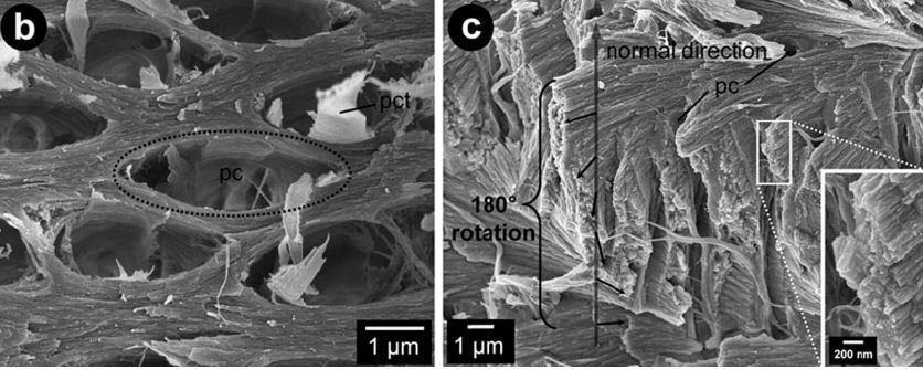
More specific this paper is about the cuticle of the lobster Homarus americanus which is a biological nanocomposite, such as most structural biological materials. It consists of a matrix of chitin-protein fibers associated with various amounts of crystalline and amorphous calcium carbonate in the rigid parts of the body, and is organized hierarchically at all length scales. One prominent design principle found in the hierarchical structure of such biological fibrous composite materials is the twisted plywood structure. In the lobster cuticle, it is formed by superimposing and gradually rotating planes of parallel aligned chitin-protein fibers. To adjust the mechanical properties to the requirements on the macroscopic level, the spatial arrangement and the grade of mineralization of the fibers can be modified. A second design principle of lobster cuticle is its honeycomb-like structure, generated by the well-developed pore canal system, whose twisted ribbon-shaped canals penetrate the cuticle perpendicular to its surface. Due to the hierarchical structure, the mechanical properties of the lobster cuticle have to be investigated at different length scales, which is essential for the understanding of the structure–mechanical function relations of mineralized tissues (e.g., potentially also bone and teeth). In order to investigate the influence of the structural principles on the mechanical properties on the
macroscopic scale miniaturized tensile, compression, and shear tests were
carried out to obtain integral mechanical data. Characterization of the
microstructure included scanning electron microscopy (SEM) combined with
energy dispersive X-ray (EDX) measurements.
Interdigitating biocalcite dendrites form a 3-D jigsaw structure in brachiopod shells
Acta Biomaterialia 7 (2011) 2237 Brachio[...]
PDF-Dokument [1.8 MB]
 Interdigitating biocalcite dendrites form a 3-D jigsaw structure in brachiopod shells
Adv. Mater. 21 (2009) 391–400 overview c[...]
PDF-Dokument [1.3 MB]
Adv. Mater. 2009, 21, 391
The cuticle of the lobster Homarus americanus is a nanocomposite, such as
most structural biological materials. It consists of a matrix of chitin-protein
fibers associated with various amounts of crystalline and amorphous calcium
carbonate in the rigid parts of the body, and is organized hierarchically at all
length scales. One prominent design principle found in the hierarchical
structure of such biological fibrous composite materials is the twisted plywood
structure. In the lobster cuticle, it is formed by superimposing and gradually rotating planes of parallel aligned chitin-protein fibers. To adjust the
mechanical properties to the requirements on the macroscopic level, the
spatial arrangement and the grade of mineralization of the fibers can be
modified. A second design principle of lobster cuticle is its honeycomb-like
structure, generated by the well-developed pore canal system, whose twisted
ribbon-shaped canals penetrate the cuticle perpendicular to its surface. Due
to the hierarchical structure, the mechanical properties of the lobster cuticle
have to be investigated at different length scales, which is essential for the
understanding of the structure–mechanical function relations of mineralized
tissues (e.g., potentially also bone and teeth). In order to investigate the
influence of the structural principles on the mechanical properties on the
macroscopic scale miniaturized tensile, compression, and shear tests were
carried out to obtain integral mechanical data. Characterization of the
microstructure included scanning electron microscopy (SEM) combined with
energy dispersive X-ray (EDX) measurements.
Adv. Mater. 2010, 22, 519–526 Multiscale[...]
PDF-Dokument [905.2 KB]
Adv. Mater. 2010, 22, 519-526
In the course of evolution nature developed materials based on organic-inorganic nanocomposites with complex, hierarchical organization from Angstroms to millimeters tailored via molecular self-assembly.Such materials possess outstanding stiffness, toughness, and strength related to their low density, while the mechanical characteristics of their underlying constituents are rather modest. This remarkable performance is a consequence of their hierarchical structure, the specific design at each level of organization, and the inherent strong heterogeneity resulting in the accommodation of macroscopic
loadings by different deformation mechanisms at different length scales. Therefore, to understand the macroscopic mechanical properties of the tissue, one should take into account
its structure–property relations at all length scales down to the molecular level. To date, this key challenge has been only partly addressed due to severe obstacles in obtaining
mechanical and structural data at the nanometer scale. The mechanical properties of important proteins and biominerals as well as some details about their exact structure are still
unknown. A powerful tool to overcome these difficulties and to better understand the structure-property relationships in biomaterials is multiscale modeling encompassing all length
scales Some progress in the development of multiscale structure-property relationships for mineralized tissues has been achieved by combined modeling and experimental approaches
applied to bone, nacre,and fish skin armor. However, these approaches do not explicitly integrate a molecular-level description and use continuum mechanics at the
meso- and macroscale (e.g., finite element analysis) coupled with experimental data obtained, for example, by nanoindentation. A more advanced multiscale modeling has been proposed
for collagen-based biomaterials, where molecular-dynamics calculations are applied to individual tropocollagen molecules, while the deformation behavior of single collagen fibrils at
the
mesoscale and of soft collagen tissues at the macroscale has been modeled by adopting continuum-mechanical concepts. First-principles calculations, based on quantum mechanics and
density functional theory (DFT), that are free of adjustable or experimental parameters have recently evolved as a versatile and accurate tool to study fundamental biological mechanisms
and structures on the atomic scale. However, due to the complexity and the hierarchical nature of biomaterials, the application of these techniques is still out of reach to study
macroscopic mechanical behavior. We therefore propose a new approach
combining ab initio calculations with hierarchical homogenization that allows us to study structure-property relations including all length scales. To describe the key concepts of this
approach and to demonstrate its applicability we choose a tissue containing chitin, the second most abundant natural polymer on earth after cellulose. Chitin-based composites serve as
exoskeleton material
for more than 90% of all animal species, a fact that demonstrates their extremely adaptive potential to realize a vast range of natural materials. In this Communication, the cuticle of a
large crustacean, the lobster Homarus americanus, is discussed as a model material. The crustacean cuticle is one of the oldest natural materials for structural and armor applications.
Crustacea, such as crab, lobster, crayfish, or shrimp, are already present in the fossil record of the Early Cambrian period (515 Myr ago) and, thus, among the most successful animals
living on earth. One of the reasons for the versatility of crustaceans is their functional body design, featuring a hard, chitin-based mineralized exoskeleton. Another consideration
for choosing the cuticle of H.
americanus is that extensive experimental data are available on its microstructure and crystallographic texture. These data are required as an input to construct the multiscale approach,
while the experimentally measured mechanical properties of the cuticle allow to quantitatively validate the accuracy of our model.